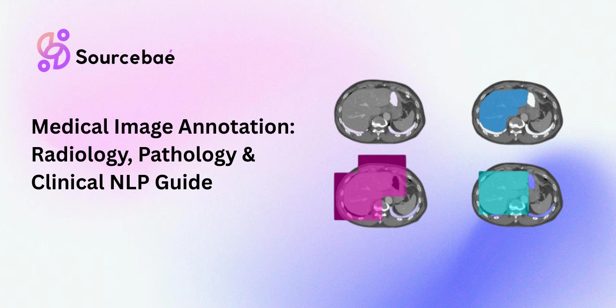

Medical image annotation is the process of labeling clinical imaging data X-rays, CT scans, MRIs, ultrasounds, and pathology slides to train AI systems that assist in diagnosis, treatment planning, and clinical research. It is the most demanding and highest-stakes domain within data annotation: the labels must be clinically accurate, the annotators must be qualified medical professionals, the data must be handled in compliance with healthcare privacy regulations, and errors carry consequences measured in patient outcomes rather than model metrics.
The scale of this domain justifies close attention. The FDA has authorized over 1,451 AI-enabled medical devices through the end of 2025, with 295 new clearances in 2025 alone and radiology imaging accounts for 76% of all authorized devices (JAMA Network Open, 2025). The global AI-enabled medical device market was valued at $13.7 billion in 2024 and is projected to exceed $255 billion by 2033 (Grand View Research). Every one of these devices depends on annotated medical imaging data for training and validation.
This post provides a comprehensive guide to medical data annotation for clinical AI: from DICOM file handling through radiology and pathology annotation workflows, clinical NLP labeling, regulatory compliance requirements, expert annotator qualifications, and quality assurance benchmarks for diagnostic accuracy.
Why Medical Annotation Needs Its Own Guide
Medical image labeling operates under constraints that do not apply to general-purpose annotation, and teams that attempt to apply standard annotation practices to clinical data consistently encounter problems that require specialized solutions.
Clinical expertise is non-negotiable.
A general-purpose annotator can be trained in a few hours to draw bounding boxes around cars in street scenes. Labeling a pulmonary nodule in a CT scan, distinguishing a benign lesion from a malignant one in a pathology slide, or identifying subtle fracture lines in an X-ray requires years of medical training. Radiologists, pathologists, and other clinical specialists are not interchangeable with crowd workers, and the cost difference reflects this: expert medical annotators command $50–$200 per hour for specialized domains, compared to general annotation rates that can be orders of magnitude lower.
Regulatory requirements govern the entire pipeline.
In general annotation, quality is a commercial concern low-quality labels lead to poor models. In medical annotation, quality is a legal and regulatory concern. The FDA evaluates the training and validation data behind every AI-enabled medical device it authorizes. The EU Medical Device Regulation (MDR) and the EU AI Act classify healthcare AI as high-risk, subjecting it to rigorous compliance requirements. Annotated medical datasets are not just training data they are regulatory evidence.
Data privacy adds operational complexity.
Medical images contain protected health information (PHI) governed by HIPAA in the United States, GDPR in Europe, and equivalent regulations globally. Every annotation workflow must include de-identification protocols, secure data handling infrastructure, and audit trails documenting who accessed what data and when. These requirements add significant overhead to project setup and ongoing operations.
The cost of errors is fundamentally different.
A mislabeled sentiment classification degrades a model’s accuracy metric by a fraction of a percentage point. A mislabeled tumor boundary in a training dataset can lead to an AI system that misses cancers or generates false positives that cause unnecessary biopsies. The error tolerance in healthcare AI training data is correspondingly tighter than in any other domain. (For foundational annotation principles that apply across domains, see our [comprehensive guide to data annotation Pillar Page].)
DICOM Annotation: Working with Medical Imaging’s Standard Format
DICOM annotation refers to the process of adding labels and structured markings to medical images stored in the DICOM (Digital Imaging and Communications in Medicine) format the internationally accepted standard for medical imaging since 1993. Understanding DICOM is essential for any team working with healthcare AI training data because virtually every imaging device in clinical use X-ray machines, CT scanners, MRI systems, ultrasound equipment produces DICOM output.
What makes DICOM different from standard image formats
A DICOM file is not simply an image. It is a structured object containing the pixel data (the image itself), patient metadata (name, ID, demographics), study metadata (referring physician, study date, imaging protocol), and series and instance identifiers that link individual images to their clinical context. This rich metadata structure is both an asset and a complexity for annotation workflows: it enables precise linking of annotations to clinical context, but it also requires careful de-identification before any annotation work begins.
DICOM annotation storage
Annotations on DICOM images are not simply drawn on the image pixels. They are stored as separate DICOM objects Structured Reports (DICOM SR) or Segmentation objects (DICOM SEG) that reference specific images through unique identifiers. This separation ensures that original clinical images remain unaltered while annotations can be updated, versioned, and shared independently.
The highdicom Python library has emerged as a key tool for bridging the ML ecosystem (NumPy arrays, Python-based model training) with DICOM-compliant annotation storage, enabling researchers to train models while maintaining clinical interoperability.
De-identification is the first step
Before any annotation begins, DICOM files must be stripped of PHI. This includes removing patient names, addresses, dates of birth, and hospital identifiers from the DICOM header attributes. Most PACS (Picture Archiving and Communication Systems) clients can export anonymized DICOM files, but for fine-grained control especially when certain metadata must be preserved for research while PHI is removed dedicated de-identification tools and protocols are necessary. Automated de-identification must also address pixel-level PHI: patient names or ID numbers sometimes appear burned into the image pixels themselves, particularly in ultrasound and older radiograph formats.
Standardization challenges
A BMC Medical Imaging study (2025) demonstrated the value of standardizing DICOM annotations by mapping body part descriptions from 116 distinct DICOM header entries to 36 standardized SNOMED CT codes, then training deep learning models to automate the classification. This kind of terminological harmonization reducing inconsistent free-text entries to controlled vocabularies is a recurring theme in DICOM annotation: the format supports rich metadata, but the metadata’s clinical value depends on standardization.
Radiology Annotation: X-ray, CT, and MRI Labeling
Radiology annotation encompasses the labeling of cross-sectional and projection imaging the workhorses of clinical diagnostics. Each modality presents distinct annotation challenges.
X-ray annotation
It involves labeling two-dimensional projection images. Common tasks include fracture detection (marking fracture lines and classifying severity), lung pathology identification (labeling nodules, opacities, pleural effusions), cardiac assessment (measuring cardiothoracic ratios), and foreign body detection. X-ray annotation is often the entry point for medical AI projects because the images are widely available, the annotation tasks are comparatively well-defined, and the imaging modality is ubiquitous in clinical practice.
CT annotation
It involves labeling volumetric data stacks of cross-sectional slices that together represent a three-dimensional view of anatomy. Annotators work across multiple slices to label structures that span many images: a lung nodule might appear across 5–10 consecutive slices, and its boundaries must be consistently traced through the volume. CT annotation commonly involves organ segmentation (labeling every pixel belonging to each organ across all slices), lesion detection and measurement (marking tumors, cysts, or abnormalities and measuring their dimensions), and vascular analysis (tracing blood vessels through the volume).
The volumetric nature of CT makes it substantially more time-consuming than X-ray annotation: a single CT study can contain hundreds of slices, each requiring individual attention. (For the foundational 2D annotation techniques bounding boxes, polygons, segmentation masks that underlie radiology labeling, see our post on [image annotation methods Post 7].)
MRI annotation
It adds complexity through its multi-sequence nature. A single MRI study typically includes multiple sequences (T1-weighted, T2-weighted, FLAIR, diffusion-weighted), each revealing different tissue characteristics. Annotators must correlate findings across sequences a lesion that is hyperintense on T2 and enhances with contrast on T1 post-gadolinium has different diagnostic implications than one that does not. This cross-sequence correlation is a form of multimodal annotation within a single imaging study, and annotation guidelines must specify which sequences should be used for delineation and how findings should be reconciled when sequences provide conflicting information.
Common radiology annotation types
It include bounding boxes (fastest to produce, used for detection tasks), pixel-level segmentation masks (most precise, used for volumetric analysis and organ delineation), landmark points (used for anatomical measurements and alignment), and classification labels (study-level or finding-level diagnostic categories). The choice depends on the downstream AI task: detection models need bounding boxes, segmentation models need pixel masks, and classification models need diagnostic labels.
Pathology Slide Annotation: Whole-Slide Imaging
Pathology slide annotation involves labeling digitized tissue samples whole-slide images (WSIs) that pathologists examine for cancer diagnosis, grading, and biomarker assessment. This is one of the most technically demanding annotation domains in all of medical AI.
The scale challenge
A single whole-slide image can be 100,000 × 100,000 pixels orders of magnitude larger than a typical photograph or even a CT slice. Annotating at this resolution means working with a pyramidal file structure that supports viewing at multiple zoom levels, from overview (entire tissue section) to high magnification (individual cells). Annotation tools must handle seamless panning and zooming across these enormous files without lag, while maintaining spatial precision for labels placed at cellular resolution.
What pathologists annotate
Common pathology annotation tasks include tumor region delineation (outlining boundaries of cancerous tissue within a tissue section), cancer grading (classifying tissue regions by severity for example, Gleason grading in prostate cancer), cellular annotation (labeling individual cells by type tumor cells, immune cells, stromal cells), biomarker scoring (quantifying expression of specific proteins, such as PD-L1 or HER2, that guide treatment decisions), and tissue type classification (distinguishing different tissue types epithelium, stroma, necrosis, fat).
Interoperability challenges in pathology
Unlike radiology, where DICOM adoption is mature, digital pathology faces what the NCI Digital Pathology Workshop (2024) described as dual interoperability challenges: syntactic interoperability (exchanging multi-resolution WSIs across different scanner vendors and proprietary formats) and semantic interoperability (harmonizing annotations, labels, and metadata across institutions).
DICOM adoption is accelerating in pathology the Slim viewer from the NCI Imaging Data Commons demonstrates DICOM-based pathology visualization but many pathology labs still work with proprietary formats (SVS, NDPI, MRXS), and annotation pipelines must handle format conversion as a routine preprocessing step.
Annotator qualifications
Pathology annotation requires board-certified pathologists or pathology trainees working under supervision. Sub-specialty expertise matters: a dermatopathologist annotates skin biopsies with different expertise than a neuropathologist annotating brain tissue. For biomarker scoring tasks (PD-L1, HER2), annotators must be calibrated against established scoring systems and inter-observer variability benchmarks.
Clinical NLP Annotation: EHR, Drug Names, and Dosages
Annotation for clinical AI extends beyond imaging to include the unstructured text that fills electronic health records (EHRs). Clinical NLP annotation involves labeling clinical notes, discharge summaries, radiology reports, and pathology reports to train models that extract structured information from free text.
Common clinical NLP tasks
It includes named entity recognition, which identifies drug names, dosages, diagnoses, procedures, and anatomical locations in clinical text. Relation extraction links entities together, such as connecting a drug to its dosage and route of administration. Temporal extraction identifies when events occurred relative to each other. For example, a patient may present with chest pain three days after starting new medication.
Negation and uncertainty detection distinguishes phrases like ‘no evidence of malignancy’ from ‘evidence of malignancy.’ This is a critical distinction that naive text models often miss. Section classification identifies which part of a clinical document a piece of text belongs to. Examples include history of present illness, physical exam, and assessment and plan.
The annotator challenge
Clinical NLP annotation requires annotators who can read and interpret medical language abbreviations, jargon, institution-specific shorthand that is often opaque to non-clinicians. “SOB” in a clinical note means “shortness of breath,” not what it might mean in general text. “WNL” means “within normal limits.” Effective annotation guidelines must include comprehensive abbreviation dictionaries and clinical context documentation.
De-identification of text
Clinical notes contain PHI in unstructured form patient names, dates, locations, and provider identifiers scattered throughout free text. Text de-identification is a distinct preprocessing step that must be completed before annotation begins, and it is more challenging than DICOM header de-identification because PHI can appear anywhere in the text in unpredictable formats.
Regulatory Compliance: FDA, EU MDR, HIPAA, and GDPR
Annotation for clinical AI is inseparable from the regulatory environment that governs medical devices and health data. Teams that treat compliance as an afterthought invariably face costly rework.
FDA requirements
The FDA evaluates AI-enabled medical devices through primarily the 510(k) pathway (97% of clearances as of 2024). The agency’s January 2025 draft guidance introduced specific transparency and labeling requirements for AI-enabled devices, recommending that manufacturers document model inputs, outputs, performance measures, and known sources of bias.
For annotation teams, this carries significant documentation requirements. Every labeled dataset used to train or validate an FDA-submitted device needs thorough process documentation. This must cover who performed the annotations and what guidelines they followed. It should also detail what quality controls were applied and how disagreements were resolved. The FDA’s Good Machine Learning Practice (GMLP) framework adds another layer. It emphasizes that training data must be representative of the intended patient population.
EU MDR and AI Act
The EU Medical Device Regulation governs medical devices marketed in Europe, and the EU AI Act (with most high-risk obligations taking effect August 2026 and full compliance for medical device AI required by August 2027) adds an additional layer. Healthcare AI is classified as high-risk under the AI Act, requiring documented data governance, bias testing, and human oversight provisions. For annotation teams, this means maintaining comprehensive records of dataset composition, annotator qualifications, quality metrics, and the measures taken to ensure the annotated data does not encode demographic biases.
HIPAA and GDPR for data handling
HIPAA (United States) and GDPR (European Union) govern how patient data is collected, stored, processed, and shared. For annotation workflows, several key requirements apply. Data must be de-identified before leaving the clinical environment. Access controls should restrict annotation to authorized individuals only. Audit trails must document every access to patient data.
Data processing agreements are needed with any third-party annotation providers. Many organizations adopt a hybrid deployment model for this purpose. Patient data stays on-premise or in a private cloud. Annotation orchestration and quality management happen in a secured cloud environment. (For broader discussion of quality frameworks and inter-annotator agreement metrics, see our posts on [annotation quality metrics Post 14] and [human-in-the-loop annotation Post 25]
Expert Annotator Requirements
The quality of medical image annotation is directly bounded by the qualifications and calibration of the annotators. This section outlines the standards that production medical annotation programs must meet.
Clinical credentialing
Radiology annotation should be performed by board-certified radiologists or radiology residents under attending supervision. Pathology annotation requires board-certified pathologists, with sub-specialty expertise matched to the tissue type and diagnostic question. Clinical NLP annotation requires clinicians or clinical informaticists who can interpret medical language in context.
Calibration protocols
Even among qualified experts, inter-observer variability is a known challenge in medicine. Two radiologists may disagree on tumor boundaries. Two pathologists may assign different grades to the same tissue section. Calibration protocols address this through several methods. Consensus sessions let annotators discuss and align on difficult cases before production begins. Reference standard sets provide gold-standard examples that annotators periodically re-annotate to check for drift. Structured adjudication sends disagreements to a senior expert who provides a consensus label.
Domain-task matching
A board-certified radiologist specializing in musculoskeletal imaging is not the right annotator for a cardiac MRI segmentation project. The annotation program must match annotators to tasks based on their specific clinical sub-specialty, not just their general credential.
QA Benchmarks for Diagnostic Accuracy
Quality assurance in medical annotation must meet higher standards than general-purpose annotation because the downstream application directly affects patient care.
Inter-annotator agreement (IAA) thresholds
For medical annotation, IAA should be measured using metrics appropriate to the task. Use the Dice coefficient for segmentation overlap, targeting ≥0.85 for well-defined structures and ≥0.70 for ambiguous boundaries. Cohen’s Kappa measures classification agreement, with a target of ≥0.75 for clinical-grade labels. The intraclass correlation coefficient (ICC) works best for continuous measurements. These thresholds should be validated against published clinical benchmarks. For example, if human radiologists achieve 0.80 Kappa on a specific task, the annotation program should match that level.
Multi-reader validation
Critical annotations should be independently produced by at least two qualified annotators, with disagreements adjudicated by a third expert. This multi-reader approach is not just a quality measure it mirrors the clinical standard of care where difficult cases receive second opinions, and it is often expected in FDA submissions.
Ongoing quality monitoring
Annotation quality is not a one-time validation it must be monitored continuously throughout production. Embedded gold-standard examples (pre-labeled cases with known correct answers inserted into the annotation queue) provide ongoing performance checks. Annotator-specific quality dashboards track individual performance over time, enabling early detection of fatigue, drift, or systematic errors.
(For a broader look at annotation quality frameworks, including IAA calculation and monitoring infrastructure, see our post on [annotation quality metrics Post 14]. Geospatial annotation shares some methodological overlap with medical imaging. For those techniques, see our post on [satellite and geospatial annotation Post 12].)
Frequently Asked Questions
What is medical image annotation?
Medical image annotation is the process of labeling clinical imaging data like X-rays, CT scans, MRIs, ultrasounds, and pathology slides. Qualified medical professionals perform this labeling to produce training and validation data for healthcare AI systems.
What is DICOM annotation?
DICOM annotation involves adding structured labels to medical images stored in the DICOM format. It uses separate DICOM objects like Structured Reports or Segmentation objects that reference specific images. This approach preserves the original clinical data fully intact.
Who can perform radiology annotation?
Radiology annotation should be performed by board-certified radiologists or radiology residents under attending supervision. Their sub-specialty expertise must match the imaging modality and clinical question.
What is pathology slide annotation?
Pathology slide annotation labels digitized whole-slide images (WSIs) of tissue samples. Key tasks include tumor delineation, cancer grading, cellular classification, and biomarker scoring. This work requires board-certified pathologists with relevant sub-specialty expertise.
What regulations govern healthcare AI training data?
In the United States, FDA requirements govern device safety and effectiveness, while HIPAA governs data privacy. In Europe, the EU MDR and EU AI Act regulate medical devices, and GDPR governs data privacy. Annotated datasets must comply with all applicable regulations.
How is quality measured in medical image labeling?
Quality is measured through inter-annotator agreement metrics like Dice coefficient for segmentation and Cohen’s Kappa for classification. Multi-reader validation with expert adjudication adds another layer of accuracy. Ongoing monitoring using embedded gold-standard examples ensures consistency over time.
What is clinical NLP annotation?
Clinical NLP annotation labels unstructured text in electronic health records. It extracts entities like drug names, diagnoses, and dosages, along with relations between them. It also captures temporal information and negation or uncertainty markers. These annotations train models that structure clinical free text.
How many FDA-authorized AI medical devices exist?
As of the end of 2025, the FDA had authorized over 1,451 AI-enabled medical devices. Radiology accounts for roughly 76% of all authorized devices. The number continues to grow, with 295 new authorizations in 2025 alone.